The ophthalmologists who took part in the Trends in Surgical and Medical Retina meeting, internationally-renowned experts in the subspeciality, stressed the importance of “small revolutions” in the operating theatre, when improving not only patient comfort, but also the prognosis.
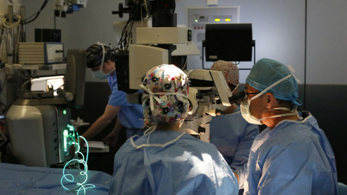
In this regard, technological innovation was highlighted as one of the key factors in the advancement of retinal surgery and occupied much of the attention of the meeting, where it was possible to view live operations featuring the use of optical coherence tomography equipment by the surgical team. Previously used just as a diagnostic tool, “applied to surgery, it can be used to obtain a cross-sectional image of the retina and enable the surgeon to see its structure with a precision that is impossible with conventional instruments, given that the tissue is extremely thin (about 200 microns)”, explains Dr Borja Corcóstegui, director of the meeting.
Regarding new ways of viewing the retina in the operating theatre, another important development was presented by the German ophthalmologist, Dr Claus Eckardt, who related his experience as a pioneer in the use of a3D system for retinal surgery. He explained that viewing a screen compared to looking through the typical microscope used today offers images of a higher quality and definition, as well as better stereopsis (depth perception), and predicted that “most colleagues will soon move away from using microscopes and start watching screens”. It does require some time to adapt, as it is a radical change in the way operations are carried out, but the ability to turn optical images into digital vision can be especially useful in difficult cases, as the surgeon is able to make adjustments to achieve excellent views.
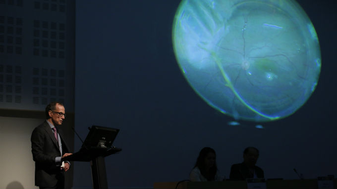
Dr. Eckardt explained his experience as a pioneer in the use of a 3D system for retinal surgery, he highlighted the advantages of converting an optical image into a digital vision, especially for handling difficult cases.
Along with this high technology, the experts also emphasised the latest advances in micro-incision surgery. The development of increasingly smaller surgical instruments makes it possible to carry out procedures with better visual, and less traumatic, results for the patient, and in half the time and, more importantly, reduce complications. In diabetic retinopathy, for example, it is now possible to reduce the formation of neovessels and postoperative inflammation, something that has also been achieved in retinal detachment surgery, according to the specialists.
Selfies of the retina
Technology has also become an ally of patients in the prevention and monitoring of chronic diseases of the retina, for which telemedicine has become a major hope for the future. According to Dr Anniken Burés, a member of the organising committee and speaker at the meeting, “one of the great challenges we face today is to find methods that will enable us to detect the disease at the moment of onset and then start treatment.
To this end, self-diagnosis and patient follow-up is key, because many retinal diseases are chronic and, to be able to control them, visits to the doctor’s surgery are required virtually every two weeks.” The optimum solution comes from one of the speciality’s great innovations, which was explained at the conference by the American ophthalmologist, Mark Blumenkranz, who presented an ambitious mobile platform that offers a number of applications to monitor the patient's visual acuity from a smartphone with different tests that make it possible to find out if there is any deformity or contrast sensitivity disorder. This way, “patients can be monitored at home weekly with highly advanced and reliable technology that makes it possible to detect minimal reactivation of the disease and apply the right treatment at the required time”, the specialist explained.
A further step is the possibility of patients taking their own retinal photographs with their smartphones, i.e. genuine “selfies of the retina”. With this in mind, Blumenkranz and his team are working on a system that, with only one lens and a plastic adapter attached to the mobile, will enable patients to take photographs of their own fundus and share them with their ophthalmologist at any time and from anywhere. “This will not only result in improved treatment and prognosis, it will also provide a lot of data from many patients around the world and enable studies to be carried out that will contribute greatly to public health.”


